Broken hearts: Shortlisted images from BHF photo competition

Winner and runners-up in the British Heart Foundation photographic competition


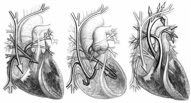
 From the heart: Heart photo competition: Blood flow
Shortlisted: The Heart's Fluid Dance: Blood flow through the heart. “We are creating computer simulations of the heart in action, in an attempt to understand better the dynamics of this entwined, sinuous dance” Dr Philip Kilner, Imperial College London
Photograph: British Heart Foundation
Photograph: Public domain
 From the heart: Heart photo competition: Cell orientation
Shortlisted: The Tree of Life: “The muscle cells of the heart intertwine like the branches of a great banyan tree. This intricate structure is essential for generating force to pump blood around the body.” The image shows the orientation of cells deep within the heart, visualised with 'multiphoton fluorescence microscopy'. Dr Patrizia Camelliti, University of Oxford
Photograph: British Heart Foundation
Photograph: Public domain
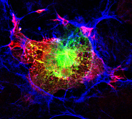
 From the heart: Heart photo competition: Fluorescent proteins
Shortlisted: COS Cell or Crab Nebula? COS cells like this one are engineered to make human forms of two proteins thought to regulate the structure of heart cells. “Staining the proteins with fluorescent antibodies allows us to see their location in the cell, and also produces images resembling something one would see from a space telescope, much less a microscope.” Joseph Dwyer, King’s College London
Photograph: British Heart Foundation
Photograph: Public domain
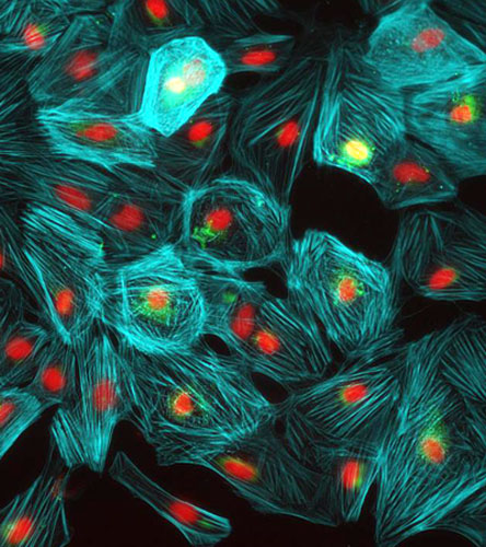
 From the heart: Heart photo competition: Heart muscle cells
Runner-up: Growing New Heart Muscle Cells. This image shows heart cells derived from human embryonic stem cells (nuclei, red; muscle protein filaments, green and blue). “Once we know more about how they grow and turn into beating heart cells, we might be able to use them to repair damaged hearts.” Dr Gabor Foldes, Prof Sian Harding, Prof Michael Schneider, Dr Nadire Ali, Imperial College, London
Photograph: British Heart Foundation
Photograph: Public domain
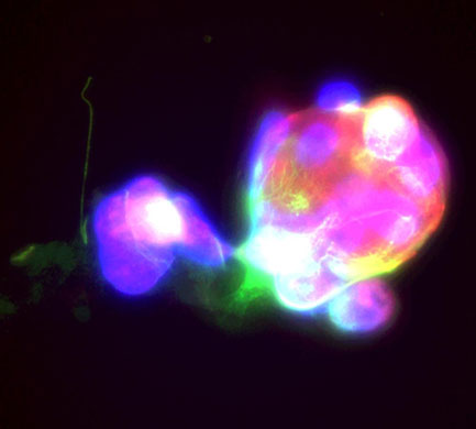
 From the heart: Heart photo competition: Megakaryocytes
Shortlisted: The Megakaryocytic Supernova. Megakaryocytes are bone marrow cells that produce blood platelets, which are vital for clotting. This image shows a megakaryocyte 'skeleton' made of F-actin (red) and tubulin (green) proteins. DNA is stained blue. “We are studying how platelets are born from megakaryocytes. In so doing, we hope to gain a better understanding of how to control the number of platelets in the blood, which could lead to new treatments for bleeding disorders.” Ms Hannah Schachtner, University of Glasgow
Photograph: British Heart Foundation
Photograph: Public domain
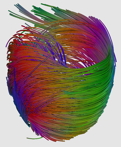
 From the heart: Heart photo competition: Muscle fibres
Runner-up: Heart Strings. This image was generated from an MRI scan of a heart using a technique called diffusion tensor imaging. The scan tracks the movement of water molecules, revealing how the muscle cells are aligned. The streamlines represent the orientation of muscle fibres in the heart’s left ventricle. 'A future application of our research would be to determine how the structure of the heart is damaged during a heart attack, and the mechanisms by which it repairs itself' Dr Patrick Hales, University of Oxford
Photograph: British Heart Foundation
Photograph: Public domain
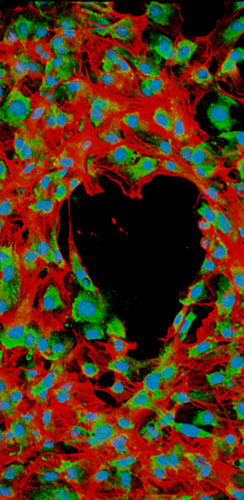
 From the heart: Heart photo competition: Inflamed artery
Winner: Looking Through the Heart. When fatty plaques are deposited in our arteries, white blood cells move from the blood into the vessel wall, and the wall becomes inflamed. The cells in this image are producing actin (red) and collagen (green). The cells’ nuclei are coloured blue. “Both actin and collagen play a key role in recruiting white blood cells when tissue becomes inflamed. A better understanding of how white blood cells interact with different components of the vessel wall will help us identify new ways to treat the inflammatory conditions that underlie cardiovascular disease.” Dr Mathieu-Benoit Voisin & Miss Doris Proebstl, William Harvey Research Institute, Barts and The London School of Medicine and Dentistry, Queen Mary, University of London
Photograph: Imaris/British Heart Foundation
Photograph: Imaris/Public domain
 

Leave a Comment

Required fields are marked *

*

*