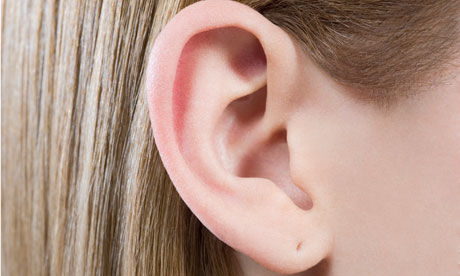
The pinna is the visible part of the ear, just right for grasping naughty boys by. Its unusual form is created in the womb, when six hillocks enlarge and unite. Mainly composed of skin and cartilage, it has lots of dips, each with its own name, such as the scaphoid fossa, triangular fossa and concha. Also known as the auricle, it works like a funnel, directing sounds into the ear canal.
People with "bat-ears" can undergo surgery to lay the pinna neatly against the head. The operation involves increasing the crease of the antihelical fold, which runs vertically down the middle of the ear and can be abnormally flat (in many hospital trusts, NHS funding for this operation has been withdrawn).
Occasionally, patients come to see me complaining of an exquisitely tender, yet hardly visible, pimple on one or other helical rim – the outermost edge of the pinna, an inflammatory condition, which if not soothed by steroid creams, may require surgical removal.
For this complaint as well as for skin cancers of the ear, a wedge excision can be performed – it is a simple operation but very satisfying. The diseased area is removed by cutting out a piece the shape of a slice of pizza, with scalpel or scissors. My personal preference is for small sharp scissors – all the layers of skin and cartilage can be cut in a neat straight line with just two firm snips. A tough non-absorbable stitch is used to bring together the two skin edges and these are left in for between a week and 10 days. The final cosmetic result is excellent and infection rates are low, thanks to the pinna's rich blood supply from arteries in the head and neck.
• Gabriel Weston is a surgeon and author of Direct Red: A Surgeon's Story

