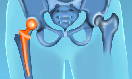
The hip joint is a ball-and-socket joint connecting the rounded head of the femur to the cup-like acetabulum of the pelvis, on each side. It has the second-largest range of movement of any joint in the body, after the shoulder. Its main function is to support us while standing, walking and running.
All babies are assessed to check that their hips are being formed in the right position. An abnormal test is one where a click is elicited by a doctor manipulating the infant's leg against the pelvis. Hip dysplasia is the name given when this has not occurred – most commonly in first-born girls. The diagnosis is confirmed by an ultrasound and the disorder treated by putting a baby in a Pavlik harness to hold their hips in the correct position. If it is not identified early enough, surgery may be necessary.
The other very common problem to affect the hip joint occurs in old age, where thinning of the bone leads to an increase in fractures of the hip, following falls. Osteoarthritis is also common, and the combination of these two explains the 50,000 hip replacements each year in the UK.
During my training, I was lucky to assist in such surgery. An incision is made down the buttock, and the top of the thigh bone is wrenched out of its socket. The head of the thigh bone is sawn off and the hip socket cleared of damaged cartilage and arthritic bone. A prosthetic ball and socket are put in place and the joint realigned before the layers of tissue overlying the joint can be repaired. All surgery can be gory, but the sounds heard in a hip-replacement operation are unforgettable.

