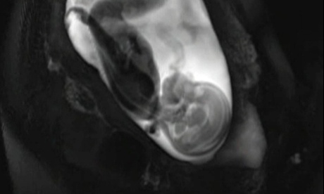
My partner is lying on her back and both of us are trying to hide our nervousness about the first ultrasound of the pregnancy. As the examination starts, we hold hands and stare intently at the bedside screen. Initially, the monitor shows little more than a shifting cauldron of grey and black organic forms but as the doctor fiddles with the machine, an image of our future son emerges from the monochrome static. I find myself looking at a picture that shows both the profile of his face and his developing brain and I am, unexpectedly, lost for words. This experience, repeated around the world, is both commonplace and astonishing. For the first time in history, most parents now see their child’s brain before they look into their eyes.
Brain development during pregnancy is key for future health, which is why it gets checked so thoroughly during prenatal examinations. But neuroscientists have become increasingly interested in how the activity of the brain becomes progressively integrated and synchronised during development to support human experience, something developmental neuroscientist Moriah Thomason calls “bringing us closer to the blueprints of the brain”. These “blueprints” are not easy to read, however, as they are encased within a tiny skull and float within the mother’s body, protected and nurtured from the outside world, making them a difficult subject for scientific study. Undeterred, groups of innovative researchers have begun to develop technology to gently and non-invasively visualise the activity of the brain as it develops in the womb.
If you’re not familiar with the challenges of neuroimaging, this is a formidable task. To complete an adult scan using functional magnetic resonance imaging, otherwise known as fMRI, stillness is essential because even slight movements can cause distortions in activity readings. To overcome this, adult participants are held in a firmly fitting head rest and software corrects for other tiny shifts in position. But as expectant mothers will tell you, the average third-trimester foetus is not an enthusiast for stillness and tends to shift and move around constantly. As a result, many neuroscientists thought that studies of the prenatal brain would be futile.
But this seemingly impossible feat has been overcome through slow, arduous manual labour. Each individual scan is like a frame from a movie that needs to be put into the same orientation. Because typical motion correction software designed for adults won’t work when scanning the foetus, aligning all of the scans has to be done by hand, for each and every individual “frame”. Thomason, an assistant professor at Wayne State University and foetal fMRI specialist, describes how it involves “more than 30 hours of work before we have the kind and quality of data that most folks using functional MRI postnatally start with”. “It is a lot of extra work,” she says, “but for very good reason.”
These reasons include some preliminary but captivating insights into the earliest stages of brain development. A recent study led by Thomason looked at how key areas begin to link up and co-ordinate their activity from week 24 of pregnancy. The researchers were able to track the emergence of wide-ranging connections that support the co-ordination of movement and information flow, where the first flickerings of synchronised activity bloom into the beginnings of the functional brain networks that remain with us into adult life.
Other studies have looked at how the foetus experiences the world outside the mother, providing a picture of an increasingly aware individual during the last trimester. Several brain imaging studies have demonstrated that in the last weeks of gestation, sounds cause clear activity in the areas of the brain involved in hearing. We know babies in late-stage pregnancy react to light but a particularly striking study, led by neuroscientist Veronika Schöpf from the University of Graz in Austria, used fMRI to determine the direction of eye gaze in the womb and showed how this was associated with surprisingly well co-ordinated neural activity in the action, visual and control areas of the brain. Many of these abilities were thought only to be present in elementary forms at birth, waiting to be shaped by experience. But we are now learning that during the last months of pregnancy, experience of the world, through the womb, is shaping the brain more fully than we previously imagined.
Although scientifically intriguing, this research area has not yet had any direct impact on medical practice, where clinicians hope to be able to better detect and treat problems during pregnancy. Catherine Limperopoulos, a specialist in foetal brain injury and neuroimaging at the Children’s National Medical Center in Washington DC, is optimistic that the basic science will lay the foundations for better care. “The potential future clinical role,” she notes, “will be to provide novel, currently unavailable insights into the neurobiological underpinnings of brain disorders” and she hopes that identifying “network specific disturbances” will be key to developing specific forms of diagnosis and treatment.
Parents won’t have the experience of staring at multicoloured measures of brain activity during their standard pregnancy checkups just yet but our understanding of the unborn brain is likely to increase vastly over the next decades. Centres for prenatal brain research are beginning to open across the world, and while we are currently impressed by the technical achievements of examining the brain during pregnancy, I suspect the biggest surprises about the earliest moments of human nature are yet to arrive.

